As many of you ask me for most of the Louis Vuitton bag prices on our Instagram
- PM (29.0 x 21.0 x 12.0 cm (length x height x width )) [Price $1,310]
- MM (31.0 x 28.5 x 17.0 cm (length x height x width) ) [Price $1,390]
- GM (39.0 x 32.0 x 19.0 cm (length x height x width )) [Price $1,470]
The Louis Vuitton Neverfull materials are Monogram, Epi Leather, Damier Ebene and Azur Canvas.
The prices for Monogram, Canvas and Damier Ebene are the same but for the Epi Leather, LV Giant and Monogram Jungle it changes. In the table below you will find the prices for every LV Neverfull bag.
NEVERFULL MM MONOGRAM JUNGLE & Giant Price: ($1,750)
NEVERFULL MM EPI LEATHER DENIM Price: $2,260
NEVERFULL MM EPI LEATHER NOIR Price: $2,090


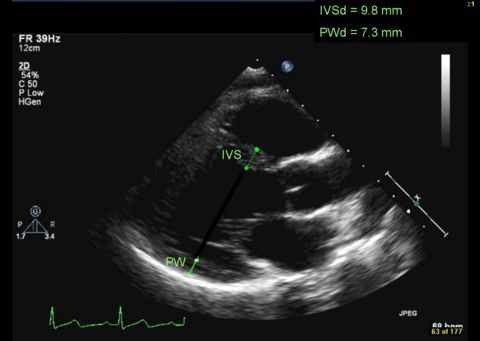
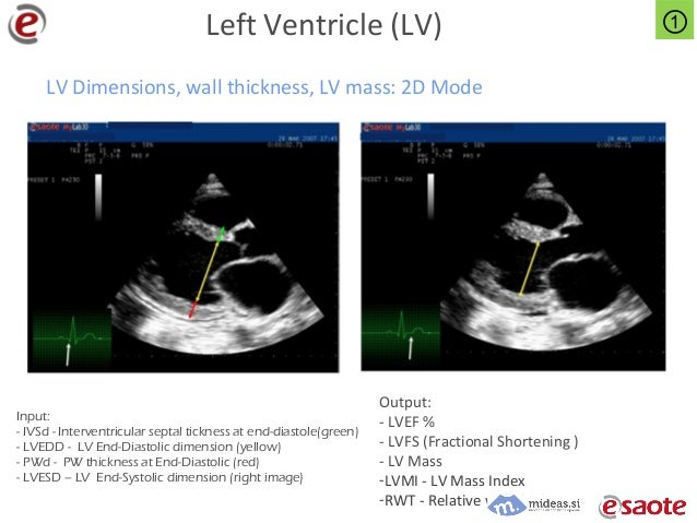
The left ventricle (LV) at the level of the papillary muscles and the right ventricle (RV) can be displayed. ... Measurements echo lv measurements should be conducted in accordance with the recommendations of the ASE, and must always be done perpendicular to the main axis of a vessel, a chamber or atria. ... The American Society of Echocardiography has published ...
How To Calculate Stroke Volume In Echocardiography (The ...
Jun 25, 2018 · Calculation For Stroke Volume In Echocardiography. If you’ve carefully obtained the above measurements, and have a reasonably modern echo machine, then you should have everything you need to obtain the left ventricular stroke volume. However, if you need to calculate stroke volume manually, then here is how you can figure it out on your own.How to Perform the Most Commonly Used Measurements in …
the left ventricle anterior wall (LVAW), the left ventricular interior diameter (LVID) and the left ventricle posterior wall (LVPW). The software is designed to perform these measurements in the following order IVS/LVAW, LVID, LVPW. Once one measurement is initiated the subsequent measurements are assumed.Measuring Left Ventricular Ejection Fraction
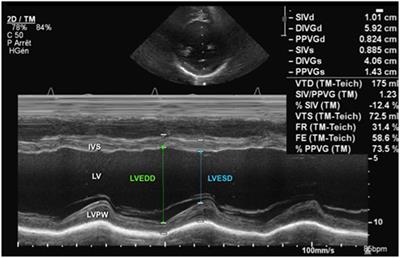
Many methods have been developed to measure LVEF using echocardiography. 1 The methods differ based on the type of echocardiographic image used (M-mode, 2D or 3D), the measurements needed and the equations/assumptions used to determine LV volumes. The measurements echo lv measurements obtained can be linear (1D), area (2D) or volume (3D) measurements.M Mode Measurements: Left Ventricular End Diastolic Volume Left Ventricular End Systolic Volume Left Ventricular Stroke volume Left Ventricular Stroke Index Left Ventricular Cardiac Output Left Ventricular Cardiac Index Ejection Fraction Left Ventricular Per Cent Fractional Shortening
While 2D echocardiography is essentially a “picture” of the heart, an M-mode echocardiogram is a “diagram” that shows how the positions of its structures change during the cardiac cycle. M-mode recordings allow in-vivo noninvasive measurement of cardiac dimensions and motion patterns of …
Echocardiography in hypertrophic cardiomyopathy diagnosis ...
Three-dimensional echocardiography (3D-E) can help in aiding diagnosis, assessing systolic function, and understanding the mechanics of SAM and LVOTO in HCM. 3D-E can provide volumetric data for accurate assessment of systolic function and has been shown to correlate well with magnetic resonance imaging for the assessment of LV volumes ...Validation of 3D Echocardiographic Assessment of Left ...
Introduction. echo lv measurements Determination of left ventricular (LV) volumes in small, young patients, especially those with complex congenital heart disease (CHD), is integral to medical and surgical management. 1,2 LV volumes have and continue to be used as key determinants of suitability for 1- versus 2-ventricular repair. 3,4 To date, measurements have been based on 2D echocardiography, applying formulas ...Introduction: Three-dimensional (3D) echocardiography has been shown to offer highly accurate measurements of left ventricular (LV) volume and mass. The present study evaluated the accuracy of 3D surface reconstruction by the piecewise smooth subdivision method in measuring volume and mass not only in the LV but also in the more complexly shaped right ventricle (RV).
RECENT POSTS:
- louis vuitton bags burlington coat factory
- best lv bag for workplace
- louis vuitton perth wa
- st louis pandora online store
- louis vuitton saintonge purse forum
- louis vuitton taurillon leather durability
- bom dia flat mule replica
- homes for sale in glenmoor country club ohio
- coach keychain wallet women
- louis vuitton handbags clutch
- adele wallet louis vuitton review
- macy's king size comforters on sale
- louis vuitton hennessy moet careers
- neiman marcus prada shoe sale