As many of you ask me for most of the Louis Vuitton bag prices on our Instagram
- PM (29.0 x 21.0 x 12.0 cm (length x height x width )) [Price $1,310]
- MM (31.0 x 28.5 x 17.0 cm (length x height x width) ) [Price $1,390]
- GM (39.0 x 32.0 x 19.0 cm (length x height x width )) [Price $1,470]
The Louis Vuitton Neverfull materials are Monogram, Epi Leather, Damier Ebene and Azur Canvas.
The prices for Monogram, Canvas and Damier Ebene are the same but for the Epi Leather, LV Giant and Monogram Jungle it changes. In the table below you will find the prices for every LV Neverfull bag.
NEVERFULL MM MONOGRAM JUNGLE & Giant Price: ($1,750)
NEVERFULL MM EPI LEATHER DENIM Price: $2,260
NEVERFULL MM EPI LEATHER NOIR Price: $2,090


Troubleshooting the ECMO circuit | Deranged Physiology
Venting the left atrium or ventricle - in the olden days one would actually access the LV apex with a cannula, and connect that cannula to the venous access lines of the ECMO circuit, allowing the ECMO pump to decompress the left ventricle. It seems to work - at least in dogs;Low-flow left ventricle percutaneous venting during ...
As a last resort, other more invasive options can directly vent the LA or LV vented via a sternotomy or thoracotomy. We elected to utilise the strategy described because the patient had a competent aortic valve that was opening with each cardiac cycle and the left ventricular end diastolic pressure was only moderately elevated.Left ventricular venting through the right subclavian ...
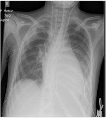
Right: Upper view of the lv vent cannula patient showing the left ventricle venting cannula and the upper limb perfusion coming from the arterial cannula inserted through the femoral artery. During ECLS, overall flow ranged from 4.5 to 4.8 l/min with a venting flow from 1.4 to 2 l/min. boy scout trash bagsNovel adjunctive use of venoarterial extracorporeal ...
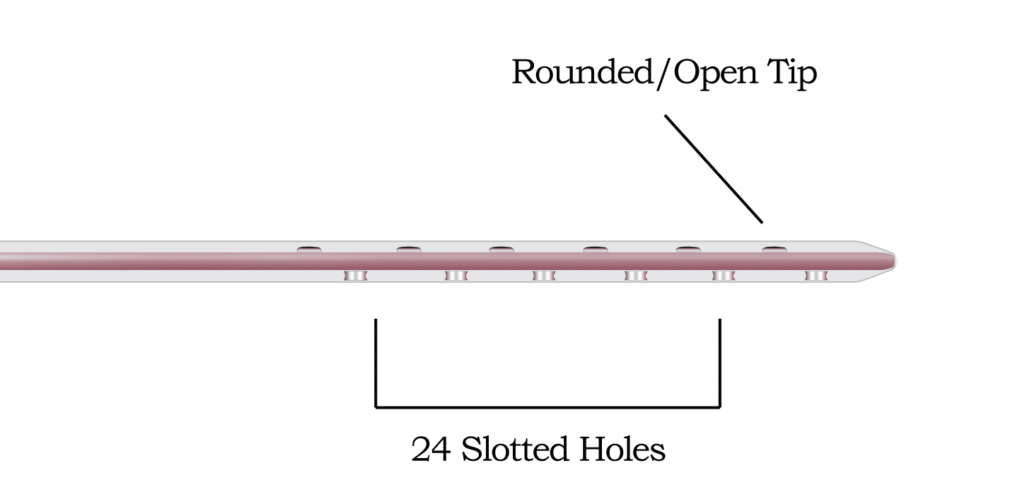
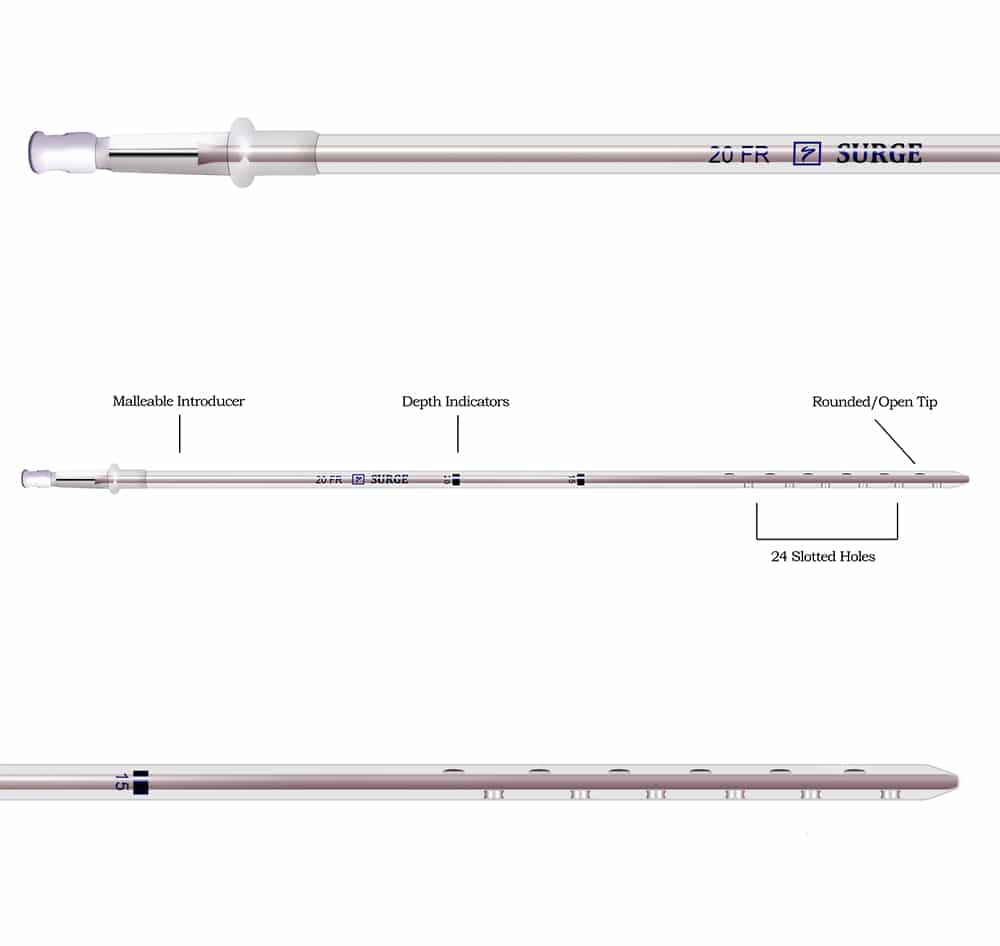
May 23, 2020 · The decision was made to convert cardiopulmonary bypass to central VA-ECMO with an LV vent . The 7.0-mm aortic cannula was left in place and the bicaval cannulae were replaced with a 34-Fr right atrial drainage cannula. To depressurize the LV, a Medtronic 22-Fr malleable venous cannula (Medtronic, Minneapolis, Minn) was inserted into LV apex ...For placement of the venous and LV vent cannula, a 6 cm right anterior thoracotomy is created lv vent cannula at the level of the fourth intercostal space, centered at the anterior axillary line. The chest is entered and the right lung is packed to prevent it from obstructing the operative field. The pericardium is identified and a pericardial cradle is ...
Sep 01, 2014 · (A) A 3/8 in Y connector is used to connect the venous cannula and the left ventricular vent to the drainage side of the ECMO circuit. Bright blood from the LV vent will mix with dark blood from the venous line. ECMO = extracorporeal membrane oxygenation; LV = left ventricular. Download : Download high-res image (465KB)
Management of left ventricular distension © The Author(s ...
decompressing lv vent cannula the left ventricle. 10. Alternatively, a percuta-neous femoral venous sheath, advanced into the left atrium using a trans-septal needle under fluoroscopic guidance, can be used as a left atrial vent incorporated into the ECMO inflow. 11. LV decompression can also be achieved by trans-septal positioning of the venous ECMO cannula ...(PDF) ECMO Cannulation Techniques - ResearchGate
“pical LV vent/cannula via left anterior thoracotomy. ... who were treated with a hybrid peripheral-central cannulation strategy accompanied by direct decompression of the left ventricle through ...A 6-8fr cannula is placed in the superficial femoral artery Spliced into arterial perfusion limb of ECMO circuit Establishes adequate perfusion of the distal leg. LV Vent Peripheral VA ECMO does not unload the LV Can actually increase LV afterload Main cause of pulmonary edema while on ECMO
RECENT POSTS:
- black gucci backpack with bee
- louis vuitton travel bags 2019-20
- alma pm damier ebene price
- louis vuitton crossbody black epi leather
- lowes black friday 2018 ad pdf
- louis vuitton receipt 2020
- gucci handbags sale outlet usa
- custom tote bag bulk
- robert kardashian louis vuitton garment bag
- lv twist wallet price
- louis vuitton store ho chi minh
- louis vuitton vernis alma bb
- louis vuitton crossbody monogram eva
- menards coupon printable