As many of you ask me for most of the Louis Vuitton bag prices on our Instagram
- PM (29.0 x 21.0 x 12.0 cm (length x height x width )) [Price $1,310]
- MM (31.0 x 28.5 x 17.0 cm (length x height x width) ) [Price $1,390]
- GM (39.0 x 32.0 x 19.0 cm (length x height x width )) [Price $1,470]
The Louis Vuitton Neverfull materials are Monogram, Epi Leather, Damier Ebene and Azur Canvas.
The prices for Monogram, Canvas and Damier Ebene are the same but for the Epi Leather, LV Giant and Monogram Jungle it changes. In the table below you will find the prices for every LV Neverfull bag.
NEVERFULL MM MONOGRAM JUNGLE & Giant Price: ($1,750)
NEVERFULL MM EPI LEATHER DENIM Price: $2,260
NEVERFULL MM EPI LEATHER NOIR Price: $2,090


Nov 15, 2011 · MRI decreases sample size requirements for detection of LV mass change by >95% (Bellenger et al. 2000), strongly suggesting that, when performing repeated measurements in training studies, MRI is a preferable technology (Myerson et al. 2002).
Magnetic resonance imaging analysis of left ventricular function in normal and spontaneously hypertensive rats 22 September 2004 | The Journal of Physiology, Vol. 513, No. 3 Comparison of fast spiral, echo planar, and fast low-angle shot MRI for cardiac volumetry at .5T
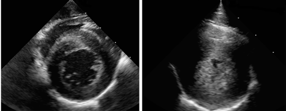
Usually, distinguishing an “athlete’s heart” from cardiopathies other than HCM is not very difficult: even when there are increases in left ventricular end-diastolic cavity dimensions exceeding the “normal range” of 53-58 mm (<3.2cm/m² in women and <3.1cm/m² in men), the absence of left ventricular systolic dysfunction is usually ...
Cardiac magnetic resonance versus transthoracic ...
Aug 18, 2009 · Cardiac magnetic resonance (CMR) is increasingly utilized for dynamic imaging of the heart with the expectation that it will provide more accurate and reproducible measurements of cardiac chamber dimensions, volumes, and function compared to other non-invasive imaging techniques such as echocardiography and nuclear cardiography [1–3].This expectation arises from the superior spatial ...Cardiac mri - SlideShare
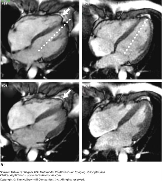
Apr 16, 2017 · The three-chamber LV (I) is obtained from a plane transecting the LV through the LV outflow tract. Normal cardiac anatomy on black-blood and bright-blood acquisitions, in sagittal; Normal cardiac anatomy on black-blood and bright-blood acquisitions, in coronal views; Normal cardiac anatomy on bright-blood two-, four- normal lv size cardiac mri and three-chamber views.Cardiac MRI | Radiology
Thus, MRI can be useful for evaluating the right ventricle in patients with suspected arrhythmogenic right ventricular cardiomyopathy (ARVC) or right-sided valvular pathology. In addition to chamber size and function, cardiac MRI has the ability to differentiate tissue characteristics (e.g. normal …Automatic Segmentation of LV and RV in Cardiac MRI
Automatic Segmentation of LV and RV in Cardiac MRI ... normal case (NOR), heart failure with infarction normal lv size cardiac mri (MINF), dilated cardiomyopathy (DCM), hypertrophic ... 256 256 by fitting maximum size of X ...Sunnybrook Cardiac Data – Cardiac Atlas Project
The Sunnybrook Cardiac Data (SCD), also known as the 2009 Cardiac MR Left Ventricle Segmentation Challenge data, consist of 45 cine-MRI images from a mixed of patients and pathologies: healthy, hypertrophy, heart failure with infarction and heart failure without infarction.Subset of this data set was first used in the automated myocardium normal lv size cardiac mri segmentation challenge from short-axis MRI, held by a ...Dr Yadav: The tranesophageal echo shows normal left ventricular size and function, normal left ventricular thickness, mild left atrial dilation, normal right atrium, normal right ventricle, and no significant valvular abnormalities. ... The key questions while performing MRI for cardiac masses are which sequence to use, which tumors are benign ... louis vuitton compact wallet mens
RECENT POSTS:
- lowepro lp36772-pww protactic camera bag 450
- coach sling backpack women's
- gucci purse pink
- who is louisa may alcott the author
- louie's pizza rehoboth beach
- house for sale on memphis nederland tx
- houses for sale in manila ut
- louis vuitton latest handbags 2020
- lv malaysia price list 2020 pdf
- louis vuitton wrapped in plastic
- louis vuitton hot stamps
- gucci baby bags outlet
- dillards sale on michael kors purses
- victorine wallet lv pink