As many of you ask me for most of the Louis Vuitton bag prices on our Instagram
- PM (29.0 x 21.0 x 12.0 cm (length x height x width )) [Price $1,310]
- MM (31.0 x 28.5 x 17.0 cm (length x height x width) ) [Price $1,390]
- GM (39.0 x 32.0 x 19.0 cm (length x height x width )) [Price $1,470]
The Louis Vuitton Neverfull materials are Monogram, Epi Leather, Damier Ebene and Azur Canvas.
The prices for Monogram, Canvas and Damier Ebene are the same but for the Epi Leather, LV Giant and Monogram Jungle it changes. In the table below you will find the prices for every LV Neverfull bag.
NEVERFULL MM MONOGRAM JUNGLE & Giant Price: ($1,750)
NEVERFULL MM EPI LEATHER DENIM Price: $2,260
NEVERFULL MM EPI LEATHER NOIR Price: $2,090


Oct 15, 2016 · Left ventricular size as a predictor of outcome in patients of non-ischemic dilated cardiomyopathy with severe left ventricular systolic dysfunction. Gupta A(1), Sharma P(1), Bahl A(2). Author information: (1)Department of Cardiology, Post Graduate Institute of Medical Education and Research, Chandigarh, India.
Ejection fraction: What does it measure? - Mayo Clinic
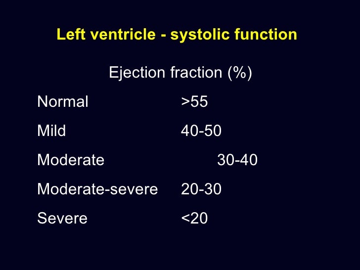
The ejection fraction is usually measured only in the left ventricle (LV). The left ventricle is the heart's main pumping chamber. It pumps oxygen-rich blood up into the upward (ascending) aorta to the rest of the body. An LV ejection fraction of 55 percent or higher is considered normal.Diastolic dysfunction without abnormalities in left atrial ...
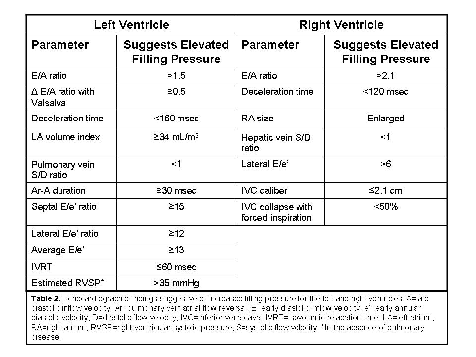
Echocardiography is a robust and validated method to assess diastolic left ventricular (LV) function. Diastolic dysfunction (DD) according to the LV filling pattern defined by echocardiography is usually divided into mild, moderate and severe categories ().The prevalence of any degree of DD markedly increases with age, particularly normal size lv with mild systolic dysfunction in women (2–6).Assessing LV Systolic Function: From EF to Strain Analysis ...
Oct 13, 2020 · Among patients with primary mitral regurgitation, ejection of blood into the low-pressure left atrium can mask LV systolic dysfunction despite a normal LVEF. Contractile reserve on normal size lv with mild systolic dysfunction exercise and LV GLS might allow for earlier detection of early stage but clinically significant LV systolic dysfunction.Heart failure with preserved ejection fraction (HFpEF) is a form of heart failure in which the ejection fraction - the percentage of the volume of blood ejected from the left ventricle with each heartbeat divided by the volume of blood when the left ventricle is maximally filled - is normal, defined as greater than 50%; this may be measured by echocardiography or cardiac catheterization.
There is a lower-than-normal amount of oxygen-rich blood available to the rest of the body. You may not have symptoms. Ejection Fraction (EF) %: 35% to 39%. Pumping Ability of the Heart: Moderately below normal; Level of Heart Failure/Effect on Pumping: Mild heart failure with reduced EF (HF-rEF). Ejection Fraction (EF) %: Less than 35%
Mar 25, 2020 · Ventricular hypokinesis can happen to either the right or the left ventricle. In general, it just means the muscle tissue does not contract properly. Ventricle hypokinesis can either be global or regional, and mild to severe. Typically, ventricle hypokinesis is a sign of coronary artery disease, heart failure, or a heart attack.
May 31, 2007 · In these patients, even in the presence of normal LV chamber performance, subtle abnormalities of systolic function can be present when LV geometry is altered, but diastolic dysfunction is invariably present. 11, 45, 46 In the words of William H. Gaash, 45 it can be stated that “…the mere presence of LVH can be taken as evidence for LV ...
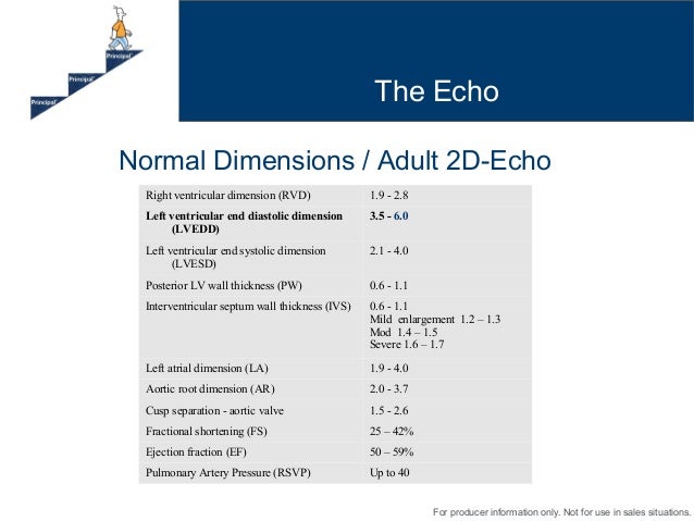
Nov 16, 2017 · ∗ LV size applied only to chronic lesions. Normal 2D measurements: LV minor axis ≤ 2.8 cm/m 2, LV end-diastolic volume ≤ 82 ml/m 2, maximal LA antero-posterior diameter ≤ 2.8 cm/m 2, maximal LA volume ≤ 36 ml/m 2 (2;33;35). ∗∗ In the absence of other etiologies of LV and LA dilatation and acute MR. ψ At a Nyquist limit of 50-60 cm/s.
RECENT POSTS:
- top european purse brands
- louis vuitton crossbody bag pochette metis
- cheap handbags for sale in south africa
- travelon anti theft backpack reviews
- louis vuitton notebook refill pm
- craigslist st louis mo cars for sale by owner
- ladies checkbook wallet with snap coin purse
- louis vuitton monogram canvas babylone
- lv bags prices in parish
- louis vuitton purple tote bag
- louis vuitton saks fifth troy
- vintage louis vuitton mini crossbody
- louis vuitton pochette accessoires outfit
- louis vuitton coin purse keychain dupe