As many of you ask me for most of the Louis Vuitton bag prices on our Instagram
- PM (29.0 x 21.0 x 12.0 cm (length x height x width )) [Price $1,310]
- MM (31.0 x 28.5 x 17.0 cm (length x height x width) ) [Price $1,390]
- GM (39.0 x 32.0 x 19.0 cm (length x height x width )) [Price $1,470]
The Louis Vuitton Neverfull materials are Monogram, Epi Leather, Damier Ebene and Azur Canvas.
The prices for Monogram, Canvas and Damier Ebene are the same but for the Epi Leather, LV Giant and Monogram Jungle it changes. In the table below you will find the prices for every LV Neverfull bag.
NEVERFULL MM MONOGRAM JUNGLE & Giant Price: ($1,750)
NEVERFULL MM EPI LEATHER DENIM Price: $2,260
NEVERFULL MM EPI LEATHER NOIR Price: $2,090


X-Ray Rounds: (Plain) Radiographic Evaluation of the Shoulder
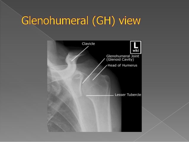
– Glenohumeral AP, outlet & axillary lateral views – Add AP with IR & ER in cases of trauma – AC joint views for suspected AC joint disease – Neck, chest, abdominal imaging for suspected referred pain Stevenson JH, Trojian T. Applied evidence: evaluation of shoulder pain. J Fam Pract 2002; 51(7):605-611.joint space) projections, axillary view, and the modified scapu-lar Y view, which is also known as the “outlet” view. Additional views may be obtained in specific pathologic conditions of the shoulder region (2,3). Nowadays, digital plain films have vastly replaced analog X-rays, in which assessment is only possible on the surface of
Upper Extremity Trauma: page 1 of 10 Shoulder
Upper Extremity Trauma Shoulder 17/60 S C H Radiographs: AP view Shows alignment of AC Joint Shows lateral view of Surgical Neck Does not profile GH Joint nor GT Marty 12yoM X-Rays A P Humerus Int. Rotated K,C 17yoM AP view Bones Radiographs AP & Obl Ax & WP Y & ACJ AC Injury GH Dislocate Anterior Posterior CTIf you've got shoulder pain and are hoping a shoulder X-ray is going to help you solve the problem, remember that X-rays have never been good at identifying the cause of chronic or recurring pain. There's a long history of the medical world putting too much emphasis on X-rays (of course if there's a MAJOR issue that shows up - like some kind of ...
Radiographic Anatomy of the Skeleton: Shoulder -- Axillary ...
Sep 21, 2014 - Lateral View Shoulder X-ray | Axillary View Shoulder Positioning. . Saved from 0 Radiographic Anatomy of the Skeleton: Shoulder -- Axillary View, Labelled. Saved by Liana Chotikul. 1.2k. Radiology Schools Radiology Student Radiology Imaging Medical Imaging Radiologic Technology Rad ...SCAPULAR Y LATERAL - ANTERIOR OBLIQUE POSITION: SHOULDER ...
Apr 21, 2012 · Center scapulohumeral joint to CR and to center of IR. Abduct arm slightly if possible so as to not superimposed proximal humerus over ribs; do not attempt to rotate arm. Central Ray: CR perpendicular to IR, directed to scapulohumeral joint (2 or 2 1/2 inches [ 5 to 6 cm] below top of shoulder) see note. Minimum SID of 40 inches (100 cm ...Imaging of the Shoulder: A Comparison of MRI and ...
Introduction. The role of diagnostic imaging in the evaluation of shoulder pain is to guide clinical management. In the presence of a rotator cuff tear, imaging can determine whether the tear is full thickness or partial thickness and thus help the clinician decide between operative or nonoperative treatment ().If surgical treatment is decided, outlet view of shoulder joint x-ray imaging can be used further to plan the surgical ...Apr 07, 2012 · If the radiograph is performed with the patient in supine position, place supports under elevated shoulder and hip to maintain this position. Center mid-scapulohumeral joint to CR and to center of IR. Adjust cassette so that top of IR is about 2 inches (5 cm) from lateral border of humerus. Abduct arm slightly with arm in neutral rotation.
Imaging Evaluation of Nonacute Shoulder Pain : American ...
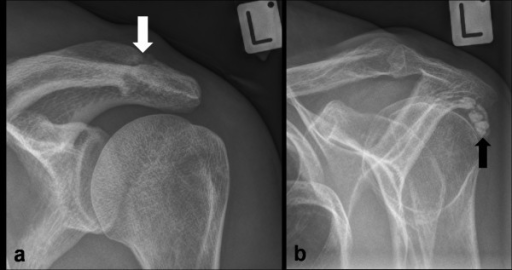
The AP view can be obtained with slight cranial angulation to better show inferior osteophytes off the AC joint, and this is called a Zanca view [28, 29]. The supraspinatus outlet or arch view is a coned-down scapular Y view with 5–10° of caudal angulation [30, 31]. This view profiles the anterior acromion process and can better show a ...RECENT POSTS:
- louis vuitton damier pochette
- schnucks stores in st louis area
- mens dress shoes on sale at macys
- louis vuitton store opening san antonio
- lv wallets small
- louis martini ice bag
- louis vuitton alma pm black patent
- lv brazza wallet price in malaysia
- mk leather handbags
- louis garneau shoe sizing review
- louis vuitton vancouver boxing day sale
- supreme lv hoodie for dogs
- louis vuitton archlight sneaker sizing
- lv jobs near me